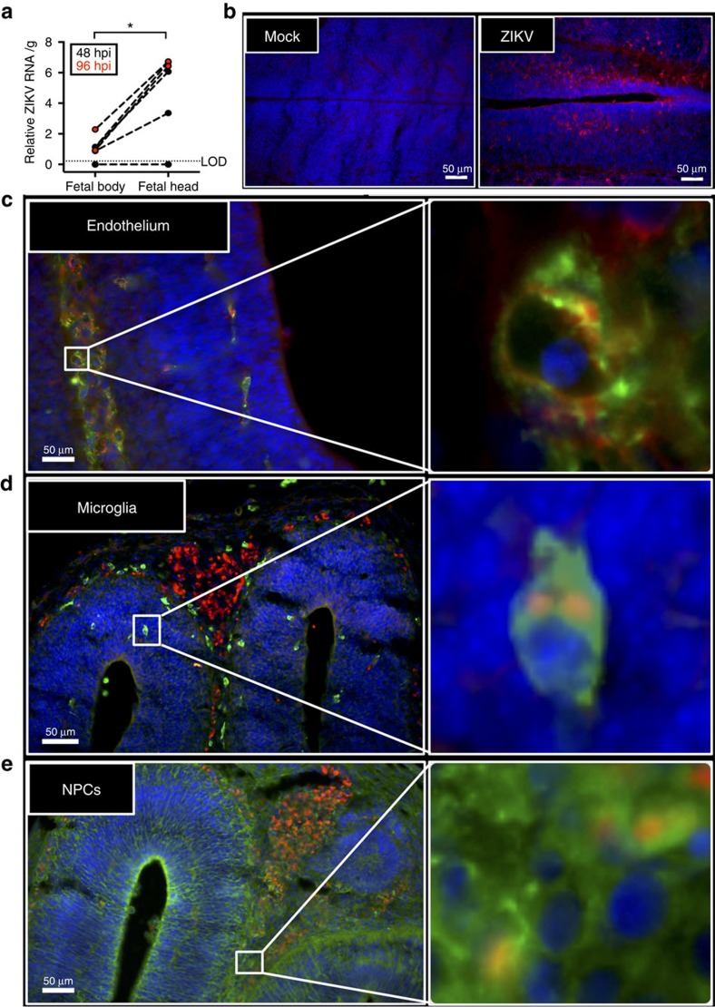
Figure 5. ZIKV antigen localizes in fetal brain cells.
Pregnant CD1 dams underwent a mini-laparotomy in the lower abdomen for intrauterine (IU) inoculation of ZIKV (1968 Nigeria) or vehicle at embryonic day 10. Fetal brains and bodies were harvested 48 or 96 h post-inoculation for quantification of viral RNA or immunohistochemical analysis. (a) ZIKV RNA was quantified in fetal bodies and heads collected 48 or 96 hpi, with the limit of detection (LOD) indicated with a dashed line. (b) Fluorescent immuno-staining of ZIKV (red) with 4′6-diamidino-2-phenylindole (DAPI, blue) to label nuclei in mock-infected (left panel) and infected (right panel) fetal brains. Scale bar, 50 μm. Fluorescent immuno-staining of ZIKV (red) with (c) CD34 (green), an endothelial cell marker, (d) Iba-1 (green), a microglial cell marker, or (e) Nestin (green), a neural stem cell marker, in fetal brains. DAPI (blue) was used to label nuclei. For c–e, the right panel is high-magnification of the white boxes in left panel. Scale bar, 50 μm. Representative images from n=10 litters are shown. For a, data were analysed with a paired t-test, *=significant difference at P<0.05.

