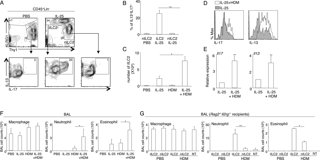
Figure 1. iILC2 are potent inducers of airway inflammation in response to acute HDM challenge.
A, C57B/6 mice were treated with PBS or IL-25 daily for 4 days. At 24 hours after the last treatment, lung hematopoietic cells were isolated and re-stimulated with PMA and ionomycin for 3 hours in the presence of monensin. Expression of IL-13 and IL-17 was examined by intracellular staining. Plots were pre-gated on CD45+Lin− cells. nILC2 were identified as CD45+Lin−KLRG1Int/loThy1hi cells. iILC2 were identified as CD45+Lin−KLRG1hiThy1lo/− cells. B, The percentage of cells that can co-produce IL-13 and IL-17 was examined. C, C57BL/6 mice were treated with IL-25 daily for 4 days, and then intranasally challenged with HDM extracts daily for 3 days. Lung iILC2 were examined by flow cytometry analysis at 24 hours after the last HDM challenge (or 96 hours after IL-25 treatment). D. Expression of IL-13 and IL-17 by iILC2 was examined by intracellular staining following PMA and ionomycin re-stimulation. Plots were pre-gated on iILC2 (CD45+Lin−KLRG1hiThy1lo/−). E. mRNA levels were examined by QPCR analysis with freshly isolated lung iILC2 without re-stimulation. Data were normalized to gapdh. F. BAL cell infiltration was examined at 24 hours after the last HDM challenge (or 96 hours after IL-25 treatment). G, 2X10⁵ iILC2 or control nILC2 were were intravenously transferred into Rag2−/− Il2rg−/− mice. Mice were intranasally challenged with PBS or HDM extracts daily for 3 days. BAL immune cell infiltration was examined at 24 hours after the last challenge. NT, no-transfer control. Data are from three independent experiments. Error bars are mean ± SEM. * P<0.05; ** P<0.01.

