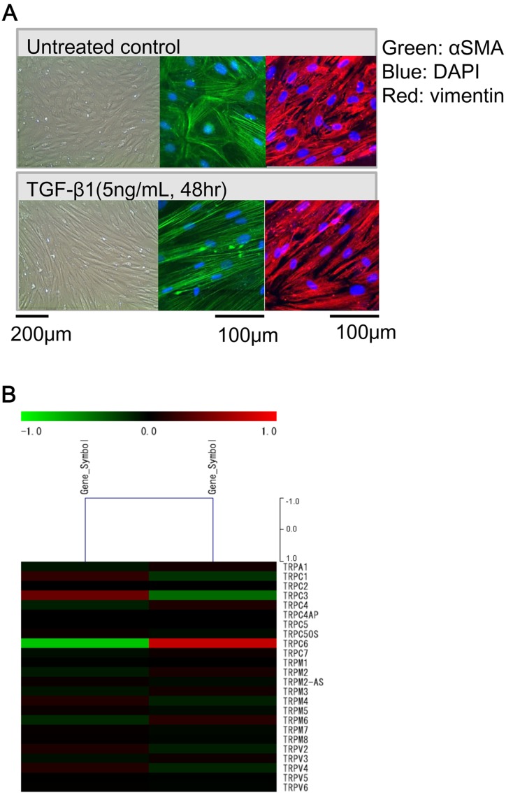
Fig. 2.
TGF-β1-induced morphological changes and TRPC6 expression in InMyoFib cells. A: Phase-contrast (left) or immunostaining images of InMyoFib cells stained with anti-α-SMA (green) and anti-vimentin (red) antibodies on the day of plating (untreated control), or 48-h post-treatment with TGF-β1 (5 ng/ml). Modified from (1) B: Fold change of TRP isoform mRNAs in InMyoFibs by expression array analysis. Control vs TGF-β1 (5 ng/ml, 24 h) treated InMyoFib.

