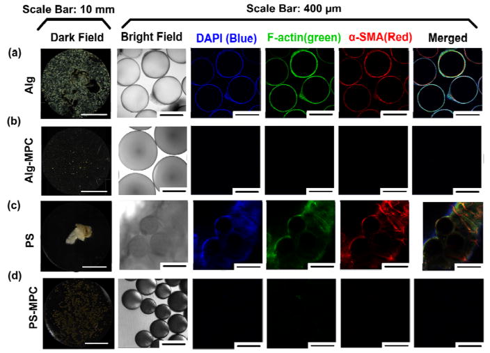
Figure 3.
Zwitterionic surface modification of microspheres reduces fibrosis in vivo. The first column (a–d) shows representative dark field microscope images for retrieved alginate and PS microspheres, 14 days after intraperitoneal implantation into C57BL/6J mice. The second column shows bright field images for stained microspheres, with their corresponding immunofluorescence confocal images (columns 3–6).

