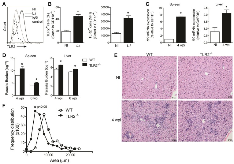
Figure 1.
TLR2 is important for host protection against L. infantum. TLR2 expression in in vitro L. infantum-infected WT BMDCs (1:5) during 24 h (A). The percentage and MFI of DC (CD11chigh) expression of TLR2 was determined by flow cytometry (B). tlr2 mRNA expression in the spleens and livers from WT mice at the 4th wpi was evaluated by real-time PCR and normalized to the constitutively expressed HPRT and GAPDH genes, respectively (C). Parasite burdens in the spleen and liver were determined in WT and TLR2−/− mice at the 4th and 6th weeks p.i. by limiting dilution (D). Representative photomicrography of stained liver sections is shown at 20 times the original magnification (E). The inflammation areas in WT (open circles) and TLR2−/− (closed circles) mice were quantified using Leica Qwin software and are shown as the frequency distribution of the lesion areas (F). Data are expressed as the mean ± SEM and are representative of three independent experiments. *P < 0.05 [Student's t-test (B,C,F) or two-way ANOVA with Tukey's post-hoc test (D)] is relative to the control group.

