Figure 3.
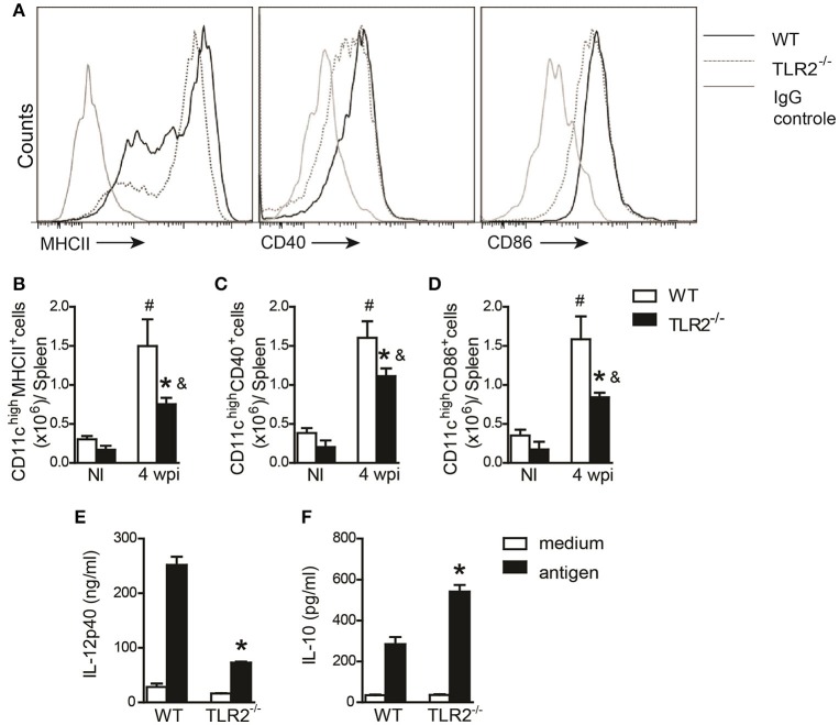
DC activation during L. infantum infection is dependent on TLR2. In vivo surface markers of DCs from uninfected (NI) and at 4th wpi WT and TLR2−/− mice were determined by flow cytometry. Histograms demonstrate the costimulatory molecules in the CD11chigh population (A) and bar graphs represent the percentage of MHCII (B), CD40 (C), and CD86 (D). All analyses were performed on CD11b+CD11chigh gated cells. WT and TLR2−/− BMDCs were infected with L. infantum (5:1) or medium for 24 h and IL-12p40 (E) and IL-10 (F) levels in the supernatants were measured by ELISA. Data are expressed as the mean ± SEM and are representative of three independent experiments, N = 4–5. *P < 0.05 compared to infected WT (B–D) or compared to stimulated WT (E,F), #P < 0.05 compared to uninfected WT, &P < 0.05 compared to uninfected TLR2−/− (two-way ANOVA with Tukey's post-hoc test).

