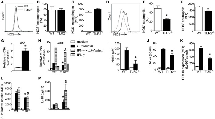
Figure 6.
TLR2 promotes nitric oxide and TNF production by neutrophils and killing of L. infantum. Spleen cells of WT and TLR2−/− mice were isolated from infected mice and stained for iNOS in CD11c−MHCII+CD11b+ cell (macrophage) and Ly6G+MHCII− cell (neutrophil) populations. Representative histograms demonstrate the iNOS expression gated in CD11c−MHCII+CD11b+ cell (A) and Ly6G+MHCII− cell (D) populations. The bar graphs represent the percentage and MFI of iNOS+ macrophages (B,C) and iNOS+ neutrophils (E,F). Neutrophils were isolated from bone marrow and infected with promastigote forms of L. infantum (5:1) or uninfected (medium). After 4 h, tlr2 (G) and inos (H) mRNA expression was analyzed by quantitative PCR, and the supernatant was collected for nitric oxide (I) and TNF (J) production, or the cells were harvested for CD11b to evaluate the neutrophil activation by flow cytometry (K). For the L. infantum uptake assay, the neutrophils were previously primed with IFN-γ (100 ng/mL) for 1 h and cultured with CFSE stained L. infantum promastigote forms (5:1) for 4 h (L) and IL-10 from the supernatant was measured (M). Data are expressed as the mean ± SEM. *P < 0.05 compared to infected WT (E,F) or WT stimulated with L. infantum (G–M), &P < 0.05 compared to WT stimulated with IFN-γ + L. infantum (H,L,M) [Student's t-test (E–G) or two-way ANOVA with Tukey's post-hoc test (H–M)] relative to the control group.

