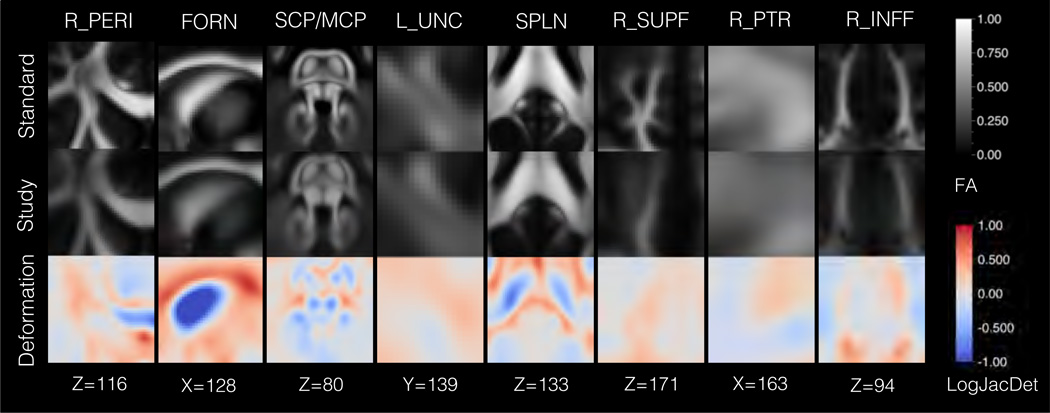
Figure 6.
Results from aging analysis in Sec. 3.2 showing the structural differences between the study and standard templates. Eight regions are shown: right anterior pericallosal white matter (R_PERI), the fornix (FORN), the superior and middle cerebellar penduncles (SCP/MCP), left uncinate fasciculus (L_UNC), splenium (SPLN), right superior frontal white matter (R_SUPF), right posterior thalamic radiation (R_PTR), and right inferior frontal white matter (R_INFF). The top row shows the standard template FA map, and the second row shows the study template FA map, which has been deformed to the standard template. The third row depicts the deformation field between the templates, with coloring to indicate the logarithm of the Jacobian determinant (LogJacDet). The LogJacDet measures the local volumetric changes induced by the deformation, where blueness indicates that contraction was required to match the standard template to the study template and redness indicates that expansion was required. The results show the greatest diiferences were in the region of the fornix, which was smaller in the study template.

