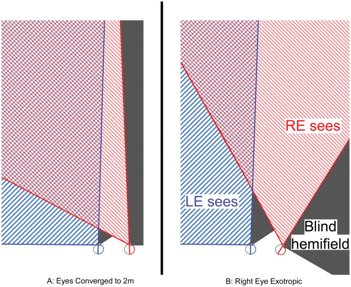
Figure 1.
A: Schematic of right hemianopia when fixating an object 2m distant (cross) and B: field expansion that occurs from right exotropia of 30°. The hashed red and blue lines represent the intact left visual fields. The areas of hemianopic vision loss are shaded gray. Field expansion comes at a cost- overlapping and double images in the portions of the field where red and blue fields overlap. Patients may or may not be bothered by double vision depending on the location of the object of interest, the type of task, and the degree of eye turn. As the magnitude of the eye turn increases, the secondary double image from the deviated eye is shifted further peripherally where it causes less interference. Therefore patients with larger angles of strabismus have more field expansion and are less likely to be bothered by the side effects of the strabismus.

