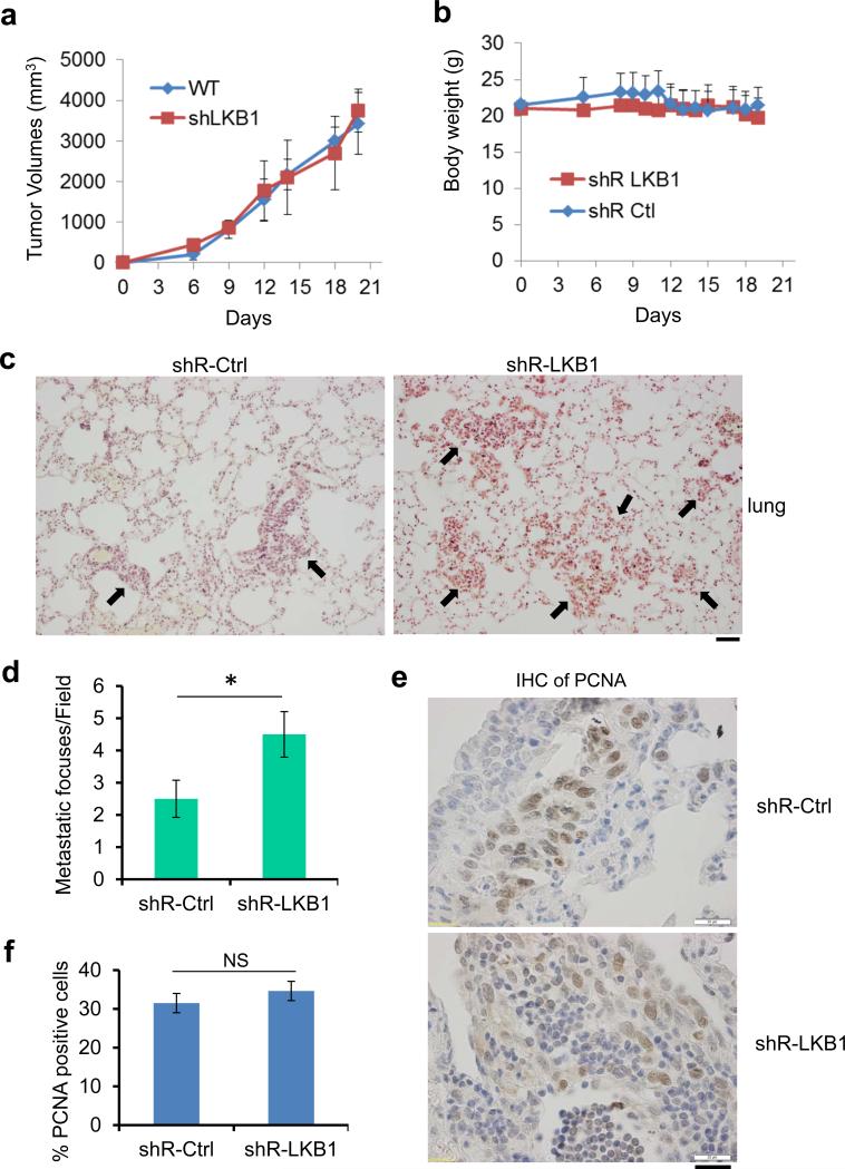
Figure 2.
Lack of LKB1 promotes lung metastasis of TC-1 cells. (a and b) 2×10⁶ TC-1/shR-Ctrl or TC-1/shR-LKB1 cells were inoculated subcutaneously into the right flank of 6 to 8 week-old female C57BL/6 mice (n=10 each). Tumor size (a) and body weight (b) of the mice were measured every three days for 3 weeks. (c and d) 2×10⁵ TC-1/shR-Ctrl or TC-1/shR-LKB1 cells were injected into each C57BL/6 mouse via tail vein (n = 10 each). Lung tumor colonies were analyzed 3 weeks post-injection. (c) Representative histological images revealed by hematoxylin and eosin staining are shown. Arrows indicate the metastatic foci. Scale bar = 50 μm. (d) Average metastatic foci in each field, at least 5 fields from each mouse were calculated (*P < 0.01, n = 10). (e) Cell proliferation marker, PCNA, was detected in the lung tumor foci with an antibody. Representative images were shown. Scale bar = 20 μm. (f) Percentage of PCNA positive cells in lung tumors with or without LKB1 expression. NS, not significant (n = 200).

