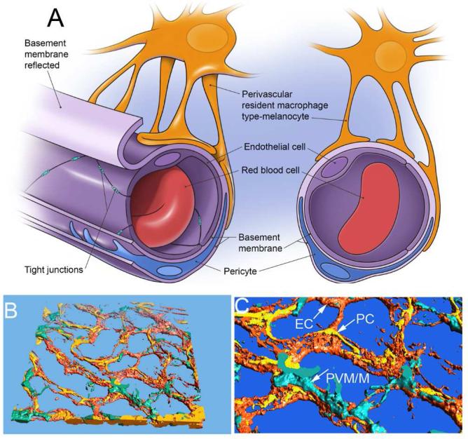
Figure 1.
(A) The illustration of a cochlear micro-vessel in cross-section shows the major components of the intrastrial fluid-blood barrier. The vessel lumen comprises ECs connected by TJs. ECs are ensheathed by a dense basement membrane shared with PCs. PVM/M end-feet cover a large portion of the capillary surface. (B) & (C) The reconstructed confocal image of the intrastrial fluid-blood barrier highlights the morphological complexity of interactions between ECs, PCs, and PVM/Ms. The PVM/Ms are immunolabeled for F4/80, PCs for desmin, and ECs with fluorescent Dil.

