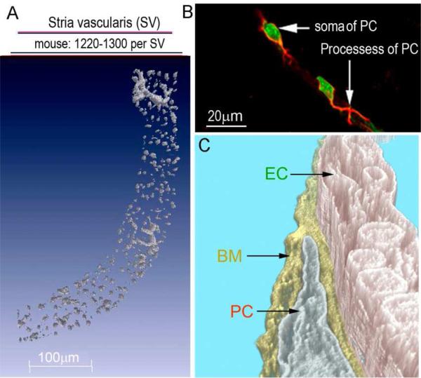
Figure 2.
(A) The super-resolution image shows the high density of strial PCs (labeled with NG2, gray) in the mouse SV (~1220-1300 PCs per SV). (B) The confocal projection shows the PC soma (stained with DAF-2DA, short arrow, green) and primary processes (labeled with desmin, long arrow, red). Two PCs are shown, each having a characteristic of a “bump on a log” shape, situated on the outer wall of a strial vessel (PC: pericyte, PVM/M: perivascular resident macrophage, V/SV: vessels of the stria vascularis, V/SL: vessels of the spiral ligament). (C) TEM tomography shows the cochlear PCs are embedded in the basement membrane (BM) and are closely associated with ECs. TEM tomography enables detection of the interactions between PCs, ECs, and the BM at high resolution (BM: basement membrane, EC: endothelial cell).

