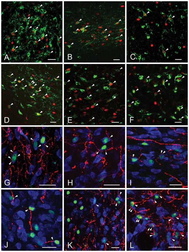
Figure 5.
Confocal images illustrate the refeeding-activated neurons (green) (A–F) and close association of PHA-L-immunoreactive (PHA-L-ir) axons (red) (G–L). The majority of refeeding-activated neurons are contacted by PHA-L-ir axon varicosities (arrowheads). The cytoplasm of neurons is labeled with HUC/D-immunoreactivity (blue). Bed nuclei of terminal stria (A, G); ventral (B, H) and lateral (I) portions of the paraventricular hypothalamic nucleus; central nucleus of amygdala (C,K); parasubthalamic nucleus (D, L); nucleus of solitary tract (E); area postrema (F) and dorsomedial hypothalamic nucleus (J).

