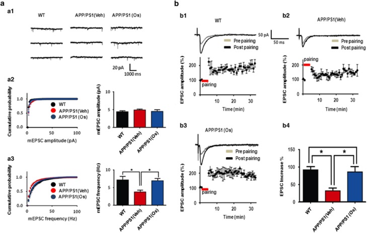
Figure 4.
Osmotin (Os) augmented the number of functional synapses without affecting synaptic strength and rescued long-term potentiation (LTP) deficits in the CA1 region of hippocampal slices from Alzheimer's disease (AD) mice. (aa1) Representative traces of spontaneous unitary excitatory postsynaptic currents (EPSCs) recorded in the CA1 region of hippocampal slices from wild-type (WT; left trace), amyloid precursor protein/presenilin 1 (APP/PS1) and osmotin-treated APP/PS1 (right trace) mice. (a2, left) Cumulative miniature EPSC (mEPSC) amplitude distributions from all recorded neurons for the three mouse groups (control (black circle), APP/PS1 (red trace) and osmotin-treated APP/PS1 (blue circle) groups). (a2, right) Summary of mEPSC amplitudes for the three groups. (a3, left) Cumulative mEPSC frequency distributions from all recorded neurons for the three groups. (a3, right) Summary of mEPSC amplitudes for the three groups. Summary of mEPSC frequencies in control APP/PS1 and osmotin-treated APP/PS1 mice. (b1, 2, 3, upper trace) A representative trace of LTP induction in the CA1 region of the hippocampi of control mice, APP/PS1 mice and osmotin-treated APP/PS1 mice, respectively. (b1, 2, 3, lower trace) Average time course of LTP induction in the CA1 region of the hippocampi of control, APP/PS1 mice and osmotin-treated APP/PS1 mice, respectively. (b4) Summary of the effects of osmotin on LTP induction in APP/PS1 mice. The bars represent the mean±s.e.m. (n=8). Significance: *P<0.05.

