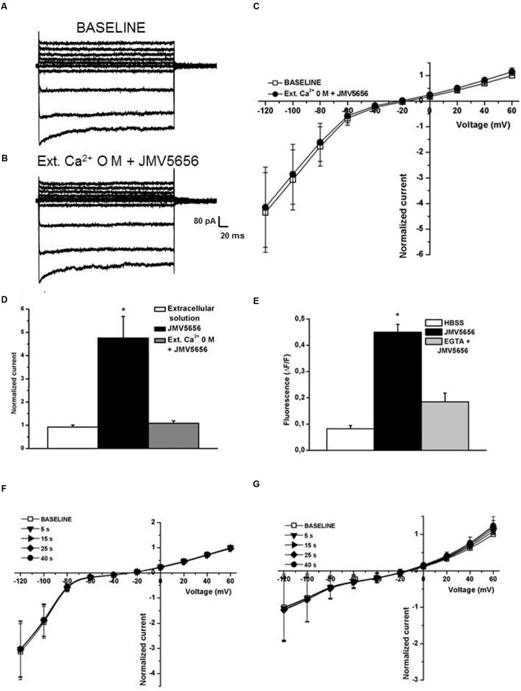
FIGURE 6.
Extracellular and intracellular Ca2+ dependence of JMV5656 effect on KCa3.1 channels. (A,B) Representative families of currents traces recorded in N9 cells perfused with the regular extracellular solution (A) and after 40 s of extracellular solution containing 0 mM Ca2+ and JMV5656 (B) in a condition of intracellular free calcium concentration of 100 nM. (C) Current/voltage relationship during perfusion with the extracellular solution (baseline, empty symbols) and perfusion containing JMV5656 (filled symbols) but lacking of Ca2+. (D) Normalized current amplitude recorded at +60 mV after 40 s of perfusion with extracellular solution (n = 11), JMV5656 (n = 9), JMV5656 without extracellular Ca2+ (n = 15). (E) Cytosolic calcium mobilization, expressed as variation in fluorescence intensity, obtained in N9 cells stimulated with the vehicle only (HBSS), with JMV5656 and with JMV5656 in presence of 1 mM EGTA to chelate the extracellular calcium. (F) Current/voltage relationship obtained in presence of JMV5656 in the extracellular solution and BAPTA 5 mM in the intracellular one (n = 5). (G) Current/voltage relationship obtained in presence of JMV5656 in the extracellular solution and 3 μM free intracellular calcium (n = 16); recording were made at selected time points (5, 15, 25, and 40 s). ∗p < 0.05 vs control group. All data have been obtained in three independent experiments.

