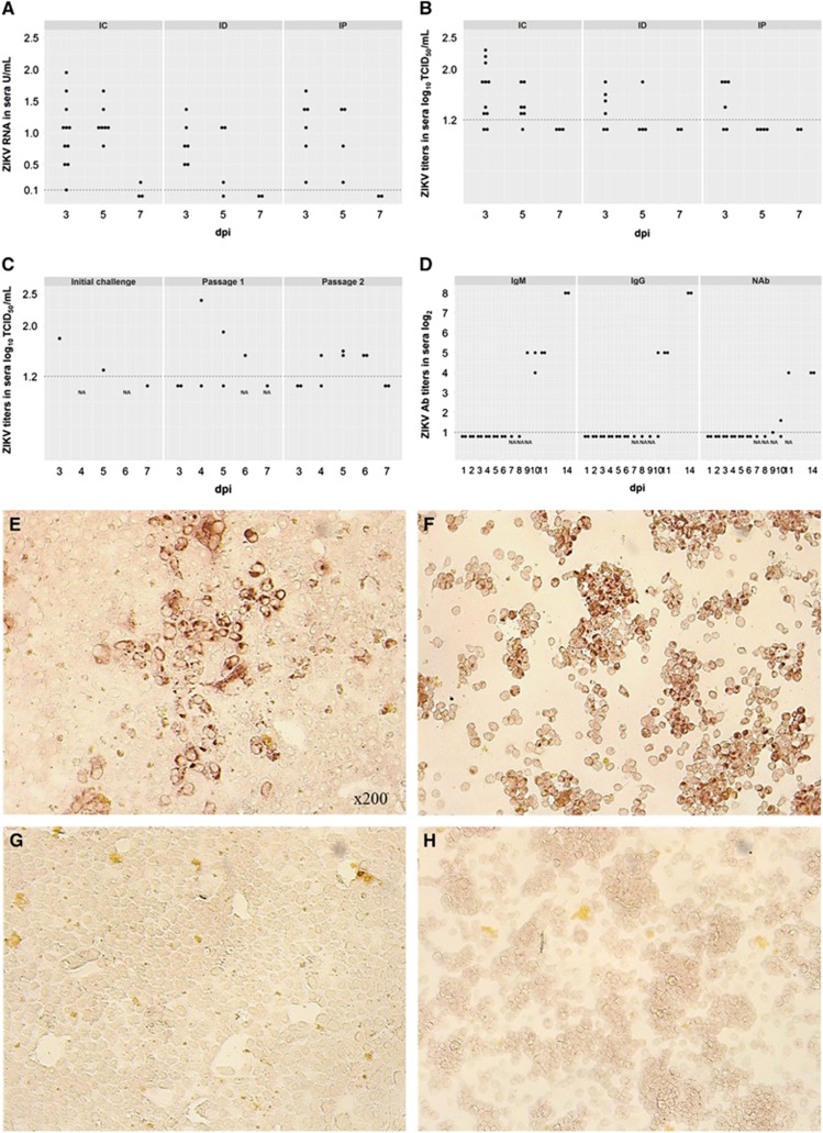
Figure 1.
Dots represent results in each sample collected from each experimental animal used in the study. A dotted line represents the assay detection limit. Dots below the detection limit are negative samples. (A) Relative ZIKV PCR titers in sera of piglets inoculated IC, ID and IP. Relative viral loads were determined using RNA from a stock of ZIKV with a known TCID50 titer. Relative log10 TCID50 values were defined as RNA units (U) and expressed as ZIKV RNA U per mL. (B) Infectious ZIKV TCID50 titers in sera of piglets inoculated IC, ID and IP. (C) ZIKV serial passage in piglets. Initial challenge – A piglet (piglet #177 Supplementary Table S1) was inoculated IC with 1 mL of inoculum containing 5.8 log10 TCID50 of ZIKV. Subsequently, serum (collected at 3 dpi) and brain tissues (collected at 7 dpi) from this piglet were used for passage 1. Passage 1 – Two newborn piglets were inoculated IC with 1 mL serum or 1 mL brain suspension (serum=1.8 log10 TCID50/mL; brain<1.2 log10 TCID50/g) from the ZIKV-infected piglet. The piglet inoculated with serum developed viremia at 5 dpi. The piglet inoculated with brain suspension developed viremia at 4 and 6 dpi. Passage 2 – Serum from passage 1 collected at 6 dpi was used to inoculate two six-day-old piglets IC. (D) Antibody titers in pigs from Passage 2. Vero E6 (E) and C6/36 (F) cells inoculated with ZIKV-positive serum and brain tissues, respectively. Vero E6 (G) and C6/36 (H) cells inoculated with samples from control pigs. The infectious ZIKV was identified by immunohistochemistry as described in Supplementary Methods. Brown staining represents active viral replication in cells. Intracerebrally, IC; intradermally, ID; intraperitoneally, IP; neutralizing antibody, NAb.

