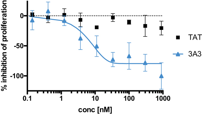
Figure 2. Cell proliferation was analysed by measuring cell growth inhibitory effects of the bivalent fusion proteins, 3A3 (filled triangles) and TAT (filled squares) incubated at concentrations ranging from 0.05 to 900 nM for 144 h with BxPC-3 cells.

Cell viability was measured using a CCK8-kit. Maximum inhibition was set to the lowest absorbance signal of 3A3 treated cells.
