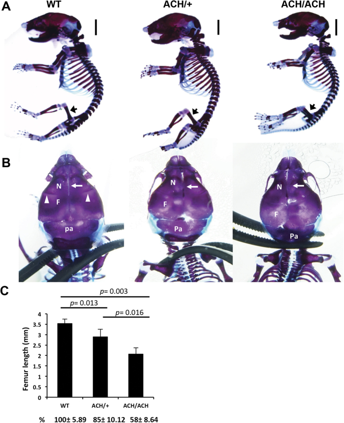
Figure 2. Skeletal defects and changes in bone architecture in newborn FGFR3ACH mice.
(A) Lateral view of skeletal preparations of FGFR3ACH/+ (ACH/+), FGFR3ACH/ACH (ACH/ACH), and WT mice. Cartilage was stained with Alcian blue and bone was stained with alizarin red. Scale bar: 30 mm. (B) Dorsal view for comparison of the skulls. The white arrowheads indicate the jugum limitans, and the white arrow indicates the metopic suture. N, nasal bones; F, frontal bones; Pa, parietal bones. (C) Femur length was significantly decreased in FGFR3ACH/ACH and FGFR3ACH/+ mice. WT, n = 3; ACH/ + , n = 6; ACH/ACH, n = 3.

