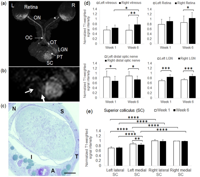
Figure 7. Mn-enhanced MRI of the visual pathway following partial optic nerve injury and binocular intravitreal Mn administration.
(a) Axial maximum intensity projection (MIP) of 3D T1-weighted MRI of the visual pathway at 6 weeks after partial transection of the right superior intraorbital optic nerve (arrowhead) and 1 day after intravitreal Mn injection to both eyes. (ON: optic nerve; OC: optic chiasm; OT: optic tract; LGN: lateral geniculate nucleus; PT: pretectum; SC: superior colliculus); (b) Enlarged MIP image at the level of the superior colliculus. Note the strong hyperintensity in the right superior colliculus projected from the left intact retina and optic nerve, in comparison to the absence of Mn enhancement in the left lateral superior colliculus (open arrow) and the mild hyperintensity in the left medial superior colliculus (solid arrow) projected from the right injured optic nerve; (c) Light micrograph of the intraorbital optic nerve section at about 1.5 mm behind the right eye at 6 weeks after partial transection of the superior optic nerve (toluidine blue stain; ×20 magnification; Scale bar = 0.1 mm) (S: superior; I: inferior; T: temporal; N: nasal; A: ophthalmic artery); (d) Quantitative assessments of Mn enhancement in the vitreous, retina, the optic nerve distal to the partial transection site, and the lateral geniculate nucleus of both hemispheres at 1 week and 6 weeks after partial optic nerve transection and 1 day after intravitreal Mn injection. T1-weighted signal intensities were normalized to the intensity in the right medial superior colliculus (Post-hoc Sidak’s multiple comparisons correction tests, *p < 0.05; **p < 0.01; ***p < 0.001); (e) Quantitative assessments of Mn enhancement in the left lateral and medial superior colliculus, and right medial and lateral superior colliculus at 1 week and 6 weeks after partial optic nerve transection and 1 day after intravitreal Mn injection. T1-weighted signal intensities were normalized to the intensity in the right medial superior colliculus. (Post-hoc Sidak’s multiple comparisons correction tests, **p < 0.01; ****p < 0.0001) (Error bars represent ± standard deviation).

