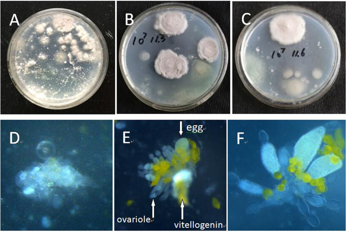
Figure 3. Fungal colony and ovary morphology of B. tabaci adult from different fungi treatment.
(A) representative images of fungal colonies derived from dissected ovaries of B. tabaci adult at 2 d age, with adult emergence from I. fumosorosea-selected B. tabaci nymph. Dissected ovary were cultured on PDA medium; (B) colony from dissected ovaries of B. tabaci adult at 5 d age, and (C) colony from dissected ovaries of B. tabaci adult at 8 d age; (D) Representative image of deformity ovary in infected insects; (E,F) images of normal B. tabaci (untreated) ovaries (ovariole, egg and vitellogenin are marked with arrows).

