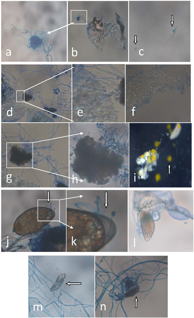
Figure 4. Representative images of I. fumosorosea growth on dissected B. tabaci fat body, egg and associated vitellogenin.
(a,b) I. fumosorosea growth on dissected B. tabaci ovaries (12 h post-incubation), (c) I. fumosorosea growth in buffer solution alone (12 h post-incubation), (d,e) I. fumosorosea growth on dissected B. tabaci fat bodies (42 h post-incubation), (f) untreated B. tabaci fat body (42 h); (g,h) effect of I. fumosorosea on associated vitellogenin (42 h post-incubation), (i) control egg/vitellogenin samples (vitellogenin marked with arrow), (j,k) I. fumosorosea growth on dissected B. tabaci eggs (42 h post-incubation), (m,n) deformation of eggs due to I. fumosorosea infection (fungal mycelia marked with arrow, 42 h post-incubation).

