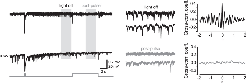
Figure 7. Optical activation of VGLUT2 neurons disrupts synchronous IPSC arrays in PCs.
LFP (top) and whole-cell current-clamp (bottom) simultaneous recordings of a control SLE triggered by a 150-ms photostimulus. Trace portions included in the shaded areas are magnified in middle panels. Right, cross-correlograms showing temporal association between IPSC arrays and LFP synchronous spikes prior to and during an 8-s lasting post-pulse.

