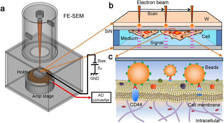
Figure 1. Overview of our dielectric microscopy of the SE-ADM system using a culture-dish holder.
(a) A schematic diagram of the SE-ADM system based on FE-SEM. The liquid-sample holder with nanoparticles and/or cultured cells is mounted on the pre-amplifier-attached stage, which is introduced into the specimen chamber. The scanning electron beam is applied to the W-coated SiN film at a low acceleration voltage. The measurement terminal under the holder detects the electrical signal through liquid specimens. (b) Overview of the liquid holder in the cultured cancer cells bound with 100-nm beads. After 4–5 days of culturing in the dish holder, the cancer cells stained with streptavidin-conjugated 100-nm beads and biotin-conjugated anti-CD44 antibodies were sealed in the bottom sample holder. The cancer cells were attached to the upper SiN film, and its W-coated side was irradiated by the scanning electron beam. (c) A conceptual diagram of the cell membrane with streptavidin-conjugated 100-nm polystyrene beads and biotin-conjugated anti-CD44 antibodies via streptavidin-biotin interaction. Biotin is not shown in the diagram.

