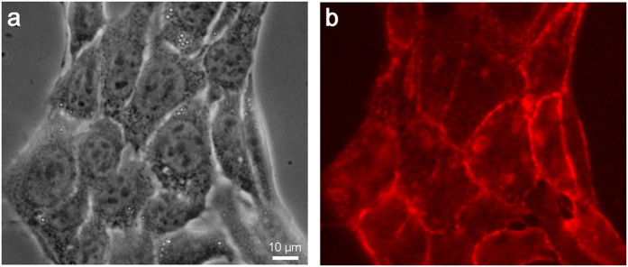
Figure 3. Optical phase-contrast and fluorescence observation images of cells stained with biotin-conjugated anti-CD44 antibodies and streptavidin-conjugated rhodamine.
(a) Optical phase-contrast image of antibody-stained cultured cells obtained with an optical microscope at 400× magnification. (b) Fluorescence image of anti-CD44 immunostained cells obtained from an optical microscope with a fluorescence filter at 400× magnification. Anti-CD44 antibodies are localized on the cell membranes. The scale bar represents 10 μm in (a).

