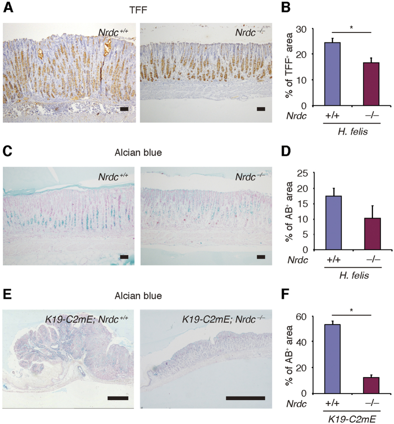
Figure 4. Metaplastic changes in Nrdc+/+ and Nrdc−/− mouse stomachs.
(A)Immunohistochemistry for TFF2 in Nrdc+/+ and Nrdc−/− mouse stomachs with Helicobacter felis infection. Bars = 100 μm. (B) Areas stained for TFF2 in Nrdc+/+ and Nrdc−/− mouse stomachs with Helicobacter felis infection. *P < 0.05. (C) Alcian blue staining of Nrdc+/+ and Nrdc−/− mouse stomachs with Helicobacter felis infection. Bars = 100 μm. (D) Areas stained with Alcian blue in Nrdc+/+ and Nrdc−/− mouse stomachs with Helicobacter felis infection. *P < 0.05. (E) Alcian blue staining of Nrdc+/+ and Nrdc−/− mouse stomachs with PGE2 expression. Bars = 1000 μm. (D) Areas stained with Alcian blue in Nrdc+/+ and Nrdc−/− mouse stomachs with forced PGE2 expression. *P < 0.05.

