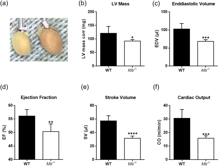
Figure 5. Mouse hearts lacking myoglobin exhibit cardiac dysfunction and atrophy.
(a) Hearts from a myoglobin deficient (Mb−/−) mouse (left) and wild-type (WT) mouse (right). (b) LV dimension–LV mass corr. (c–f) Systolic functional measures–end-diastolic volume (EDV), left ventricular (LV) ejection fraction (EF), stroke volume (SV), cardiac output (CO) using echocardiography in WT and Mb−/− mouse hearts. Values are mean ± SD, p < 0.05, n = 5.

