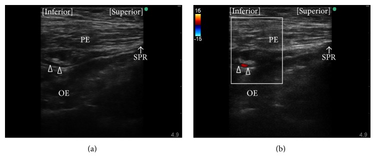
Figure 4.
Observation of the obturator artery. (a) Long axis view of a luminal structure (open triangles) is seen at the plane between the pectineus and obturator externus muscles during preprocedure observation using the approach described by Akkaya et al. [35]. (b) The luminal structure was confirmed to be the obturator artery using color flow Doppler. PE, pectineus muscle; OE, obturator externus muscle; SPR, superior pubic ramus.

