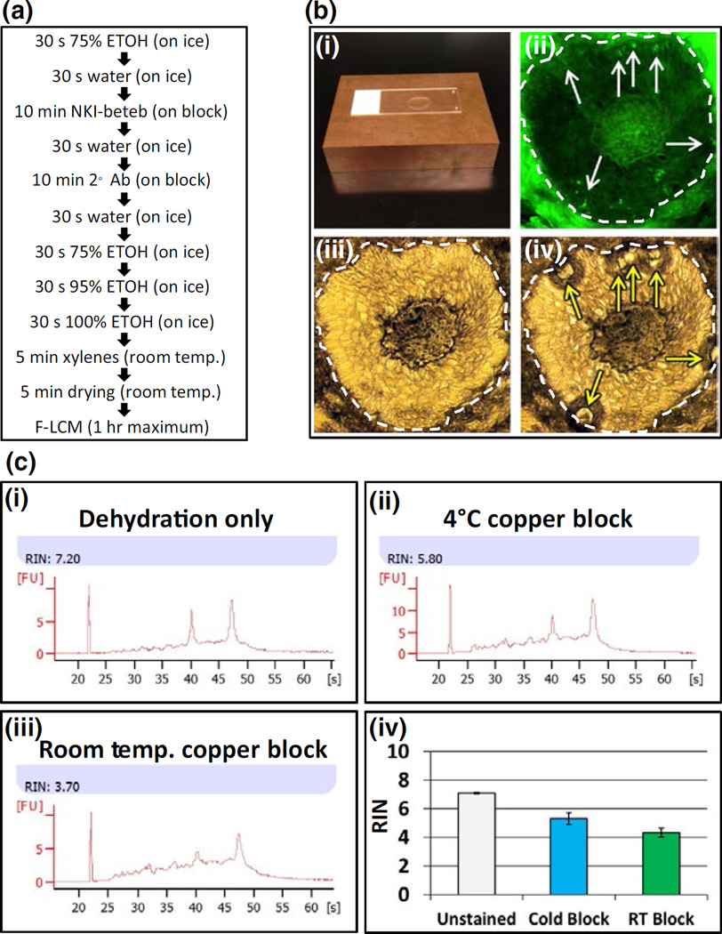
FIGURE 1.
Isolation of individual melanocytes (MCs) from epidermis and hair follicles. Panel a: Rapid immunostaining protocol to preserve RNA quality using NKI-beteb antibody. Panel b: The copper blocks used for rapid immunostaining at 4°C to preserve RNA integrity (b-i); NKI-beteb- positive MCs (fluorescent field, b-ii white arrows) laser captured after immunostaining. Bright field images show sample before (b-iii) and after (b-iv) laser capture indicating spots where tissue was captured (yellow arrows). Panels ii, iii and iv, scale bar: 50 µm. Panel c: RNA integrity of tissue scraped from slides using different staining methods. Slides stained using cold copper block (c-ii; c-iv, blue bar) showed less degradation than slides stained at room temperature (c-iii; c-iv, green bar); unstained slides showed the best RNA integrity (c-i; c-iv, white bar).

