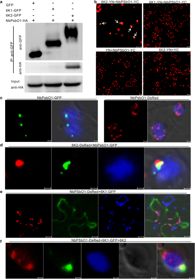
Figure 3. Interaction of 6K2 with NbPsbO1.
(a) Co-immunoprecipitation (Co-IP) assay for 6K2 and NbPsbO1. 6K2-GFP and NbPsbO1-HA were co-expressed in N. benthamiana leaves via agro-infiltration. N. benthamiana leaves co-expressing 6K1-GFP and NbPsbO1-HA, or GFP and NbPsbO1-HA were used as negative controls. (b) Images of N. benthamiana cells co-expressing 6K2-YN and NbPsbO1-YC, 6K1-YN and NbPsbO1-YC, YN and NbPsbO1-YC, 6K2-YN and YC. Punctates containing 6K2-YN and NbPsbO1-YC were indicated with arrows. (c) Transient expression of NbPsbO1-GFP and NbPsbO1-DsRed in N. benthamiana leaf epidermal cells. Images (left to right) show NbPsbO1-GFP green punctates, overlay of NbPsbO1-GFP green punctates with chloroplast auto-fluorescence, NbPsbO1-DsRed red punctates, and overlay of NbPsbO1-DsRed red punctates with chloroplast auto-fluorescence, respectively. (d) Co-localization of 6K2-DsRed and NbPsbO1-GFP in association with a chloroplast. Images (left to right) show 6K2-DsRed red punctate, NbPsbO1-GFP green punctate, chloroplast auto-fluorescence, and overlay of the three images. (e) Localization of NbPsbO1-DsRed punctate and 6K1-GFP fluorescence in a cell. Images (left to right) show NbPsbO1-DsRed red punctate, 6K1-GFP fluorescence, chloroplast auto-fluorescence, and overlay of the three images. (f) Co-expression of NbPsbO1-DsRed, 6K1-GFP and non-tagged 6K2 in a cell. Images (left to right) show NbPsbO1-DsRed fluorescence, 6K1-GFP fluorescence, chloroplast auto-fluorescence, differential interference contrast and overlay of the four images. All images were taken at 48 hpai. Scale bars in (c), (d and f) equal to 1 μm; Scale bars in others panels equal to 10 μm.

