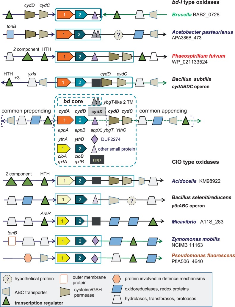
Fig. 2.—
Graphical representation of the characteristics of the gene clusters for bd oxidases. The basic structure of the gene clusters for bd oxidases conforms to those of cydABDC (top) and ythABC (bottom) operons found in Bacillus organisms (Winstedt et al. 1998; Winstedt and von Wachenfeldt 2000). The gene accession for cydA is reported for several taxa. Diverse graphical symbols indicate functionally different proteins, as illustrated in the legend at the bottom of the figure. The various names for gene annotation of the core subunits of bd oxidases are listed within the central dashed square. Structurally different forms of the small hydrophobic subunit cydX are represented as follows: Two adjacent triangles, two transmembrane segment-proteins of acetic acid and other bacteria; a triangle, the single transmembrane cydX proteins of bd-I type oxidases; a rhombic, the single transmembrane DUF2474 proteins. A black square indicates the apparent absence of this subunit (gap) in the gene cluster.

