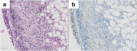
Fig. 4.

Example of an adipose lesion negative for HMGA2 immunostaining (case 6). This image was obtained from biopsies of a bronchial lesion located in the left lower lobe. Hematoxylin and eosin staining (a) and HMGA2 immunostaining counterstained by hematoxylin (b). The lesion is located in the lamina propria and consists of sheets of adipose cells. The nucleus of adipose cells negative for HMGA2 immunostaining. In contrast, some nuclei of superficial epithelial cells were positive. Scale bar 50 μm
