Fig. 2.

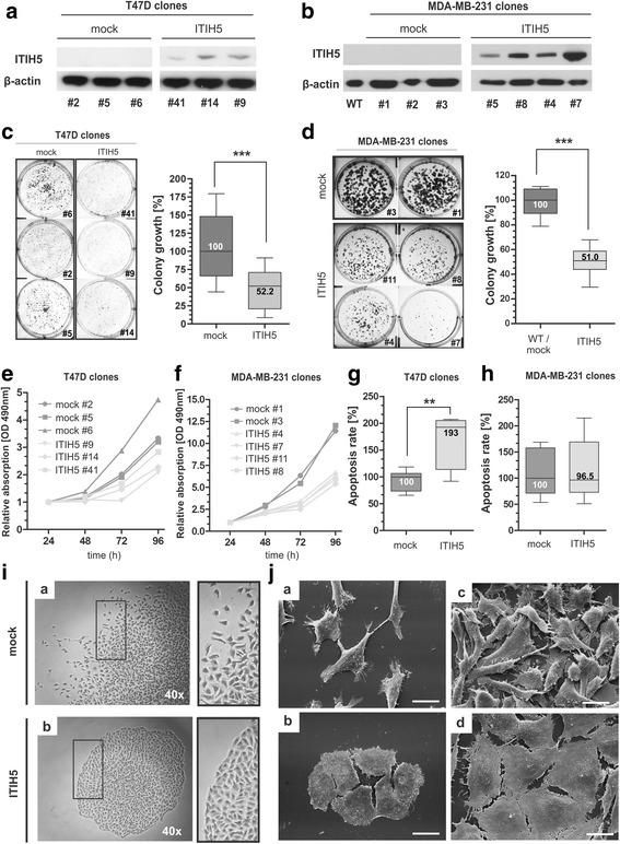
ITIH5 impairs cell growth and colonization of breast cancer cells and induce a phenotype shift in vitro. a ITIH5 gain-of-function model of luminal breast cancer cells: Ectopic ITIH5 expression in transfected T47D ΔpBK-ITIH5 clones was confirmed by Western blotting. A specific signal of the ectopic ITIH5 protein is detectable only in T47D ITIH5 clones. β-actin served as loading control. b ITIH5 gain-of-function model of basal-type breast cancer cells: Ectopic ITIH5 expression in transfected MDA-MD-231 ΔpBK-ITIH5 single-cell clones was confirmed by Western blotting. A specific signal of the ectopic ITIH5 protein is detectable in MDA-MB-231 ITIH5 clones. β-actin served as loading control. c Colony growth of luminal T47D breast cancer cells in dependency of ITIH5 re-expression. Box plot presents averages of triplicate experiments based on three independent T47D ITIH5 and three T47D mock clones. Left: Representative wells with grown ITIH5 as well as mock colonies are shown. Right: Densitometrical evaluation of colony growth after 14 days. d Colony growth of basal-type MDA-MB-231 breast cancer cells due to stable ITIH5 re-expression. Box plot presents averages of triplicate experiments based on four independent MDA-MB-231 ITIH5 and two MDA-MB-231 mock clones. Left: Representative wells with grown ΔpBK-ITIH5 as well as mock colonies are shown. Right: Densitometrical evaluation of colony growth after 14 days. e-f XTT proliferation assay was performed. T47D e and MDA-MB-231 f ITIH5 single-cell clones showed reduced cell growth compared with ΔpBK-mock controls. The baseline level at 24 h was set to 1. g-h Caspase 3/7 activity as indicator of apoptosis in independent T47D g and MDA-MB-231 h mock and ITIH5 single-cell clones (n = 3, respectively). Box plot demonstrates relative apoptosis rate. Horizontal lines: grouped medians. Boxes: 25–75% quartiles. Vertical lines: range, minimum and maximum, **p < 0.01. i Comparison of morphological MDA-MB-231 colony growth patterns of ITIH5 and mock clones. Right images: colony edges. Representative light-micrographs are shown. j Comparison of single-cell plasticity showing different confluence of both MDA-MB-231 ITIH5 and mock clones. Representative SEM-micrographs are shown. Scale bar = 20 μm
