Fig. 3.

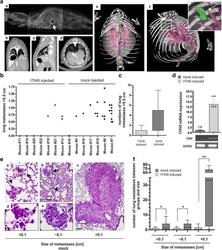
ITIH5 suppresses lung colonization of basal-type breast cancer cells in vivo. a In vivo μCT screening highlighted metastatic growth in mouse lungs. Representative 2D (a–d) and 3D (e + f) images of the lung after contrast-agent application and 3D volume rendering are shown. Macro-metastases foci (white circles; green-colored after segmentation) in mice intravenously injected with MDA-MB-231 mock cells (control set) in the pleural space. Red: vascular structures. Blue: tracheobronchial system. b Quantification of metastases by in vivo μCT analyses: Number and nodules size of lung metastases for each mice (n = 7) of the ITIH5 set (ITIH5 clones) compared with the control set (n = 7) is illustrated. c Box plot illustrating reduced numbers of grown metastases in mice injected with MDA-MB-231 ITIH5 cells. d Human ITIH5 mRNA in ITIH5-induced lung tumors compared with ΔpBK-mock-induced tumors. Columns: Mean of triplicate determinations. Error bars, + standard error of margin (s.e.m.). e Representative H&E stained metastases of each size category of mock-treated animals. Black arrows: tumor nodules. Framed rectangle regions are separately enlarged. Scale bar: 100 μm. f Box plot grouped by three metastases size categories verified a decrease of metastasis growth in mice injected with MDA-MB-231-ITIH5 cells (n = 7) compared with mice of the control set (n = 7), p < 0.05, **p < 0.01
