Fig. 8.

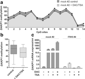
In vitro demethylation of the DAPK1 5’UTR locus correlates with DAPK1 re-expression in ΔpBK-mock cells. a Pyrosequencing analysis for each CpG dinucleotide (1–14) within the DAPK1 5’UTR region determined prior (−DAC/-TSA; dark-grey-filled) and after in vitro demethylation treatment (+DAC/+TSA; grey-filled). b Box plot analysis shows reduction of the median methylation ratio within the DAPK1 5’UTR region in ΔpBK-mock cells after DAC/TSA treatment (+) compared to non-treated cells (control). Horizontal lines: grouped medians. Boxes: 25–75% quartiles. Vertical lines: range, peak and minimum; **p < 0.01. c Real-time PCR results illustrate a clear DAPK1 re-expression after treatment with both DAC and TSA (+) in mock clones while now further expression of DAPK1 mRNA was detected in ITIH5 clones already harboring an unmethylated DAPK1 5’UTR region. Non-treated cells (-DAC,-TSA) were set to 1, respectively. Error bars: + s.e.m
