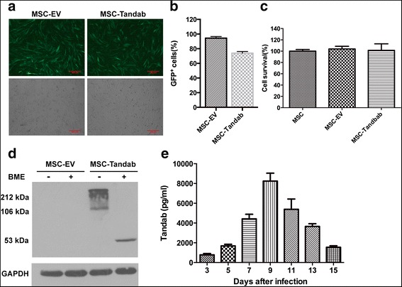
Fig. 2.

Constitutive expression of Tandab (CD3/CD19) in MSCs. MSCs were transduced with lentivirus coding Tandab (CD3/CD19) at 8 MOI for 12 h. Then, supernatants were removed, and fresh medium culture was added. a The representative images depicted the infection efficiency of MSCs with lentivirus. Forty-eight hours after infection, MSCs carrying copGFP were observed under fluorescent field (upper panel) and bright field (lower panel), scale bar = μm. MSC-Tandab, MSCs transduced with lentivirus coding Tandab (CD3/CD19); MSC-EV, MSCs transduced with empty lentivirus. b FACS analysis of percentages of copGFP-positive MSCs. c Cell survival of MSCs transduced with or without lentivirus were detected by the MTT assay. d Western blot analysis was employed to determine the protein expression of Tandab (CD3/CD19) in MSCs with anti-His tag antibodies after 5 days of transduction. GAPDH, served as a loading control; BME, β-mercaptoethanol. e Transduced MSCs secreted Tandab (CD3/CD19) constantly. MSC-Tandab and MSC-EV were cultured in a 24-well plate (4 × 10⁴/well). And the level of Tandab (CD3/CD19) released into culture was measured by ELISA in the indicated time. Data shown are the mean ± SD of the three repeated experiments
