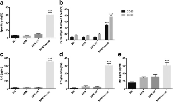
Fig. 4.

Cytotoxicity of T cells to Raji cells mediated by Tandab (CD3/CD19) secreted from MSCs. In order to evaluate the function of MSCs-secreting Tandab (CD3/CD19), a co-culture system using transwell plates with 0.4-μm-pore membrane was established. MSCs were plated into 6-well plates with a density of 1 × 10⁵ cells per well after transduced with lentivirus. Seventy-two hours later, Raji cells were labeled with calcein-AM (5 μM). Then, PBMCs and labeled Raji cells (E:T=10:1) were added to the equilibrated inserts. After co-cultured for 24 h, cells in the inserts were harvested to be detected by FACS. a The specific lysis of Raji cells. b Activation surface markers CD69 and CD25 of T cells. c–e Cytokines including IL-2, IFN-γ, and TNF-α in the supernatant were measured using ELISA kits. PR, PBMC + Raji; MPR, MSC + PBMC+Raji; MPR-EV, MSC-EV + PBMC+Raji; MPR-Tandab, MSC-Tandab + PBMC + Raji. ***P< 0.001 compared with PR group. Data shown are the mean ± SD of the three repeated experiments
