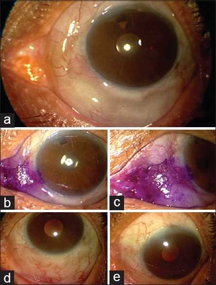
Figure 1.

(a) Top row – slit-lamp photograph of left eye (at presentation) showing a diffuse high bleb spreading nasally and inferiorly along the limbus. (b and c) Middle row – preparation of eye for laser photocoagulation, by staining with gentian violet. Gentian violet has been shown to enhance laser penetration. (d and e) Bottom row – slit-lamp photograph of left eye (6-week postintervention) showing limitation of spread of bleb nasally as well as inferiorly
