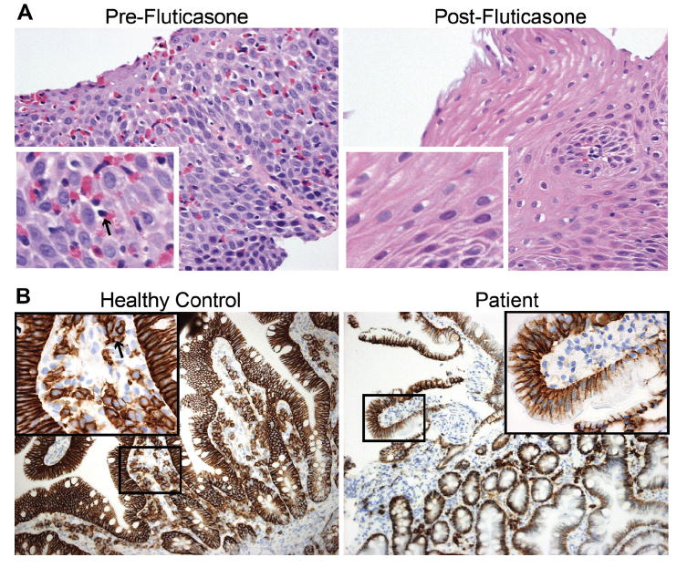
FIGURE 2.

A, Esophageal biopsy showing eosinophils (example marked with an arrow in the “Pre-Fluticasone” inset) in mucosa before but not after fluticasone therapy. Large images at 400×, insets at 1600×. B, Duodenal biopsy from non—common variable immunodeficiency (CVID) control (left) and patient (right) with immunohistochemistry staining of CD138+ cells (example marked with an arrow in the “Healthy Control” inset) demonstrating the absence of plasma cells in the patient’s gastrointestinal (GI) tract. Large images at 200×. Zoomed-in areas are 400× and correspond to the box in the low magnification image.
