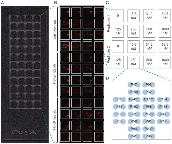
Fig. 2.
Optimization of a G9a KMT assay on microarrays. (A) A 48-well optimization array partitioned by hydrophobic wax. (B and C) Scanned image of an optimization array assay following a 2-h incubation with seven G9a concentrations, a no enzyme control (0), and three detection antibodies (H3K9me1, EpiCypher #13-0014; H3K9me2, Abcam #1220; H3K9me3, Active Motif #39161), all in duplicate. (D) Print layout of individual arrays partitioned on 48-well optimization slides. Each array consists of 24 unique histone peptides printed in duplicate. Peptide identifications numbers correspond to those used previously (Rothbart et al., 2015). P245 and P177 are H3(1–32) and H3(1–43), respectively. Biotin-4-Fluorescein (not shown) is printed in the corners of each array as a spotting control and a landmark to aid in alignment for data analysis.

