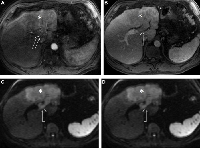
Figure 3.
HCC in a 67-year-old man with alcoholic cirrhosis.
Notes: Axial post-contrast T1-weighted MR images in arterial (A), delayed phase (B), axial T2-weighted (C), and diffusion weighted (D) MR images demonstrate an infiltrative mass (asterisk) with ill-defined margins, exhibiting heterogeneous enhancement in the arterial phase, washout in the delayed phase with moderately increased signal intensity on T2-weighted and diffusion restriction on diffusion-weighted images. There is soft tissue noted within the left portal vein (arrow) exhibiting all signal characteristics and contrast enhancement similar to the tumor, representing LR-5V. See Table 2 for LI-RADS classifications.
Abbreviations: HCC, hepatocellular carcinoma; MR, magnetic resonance.

