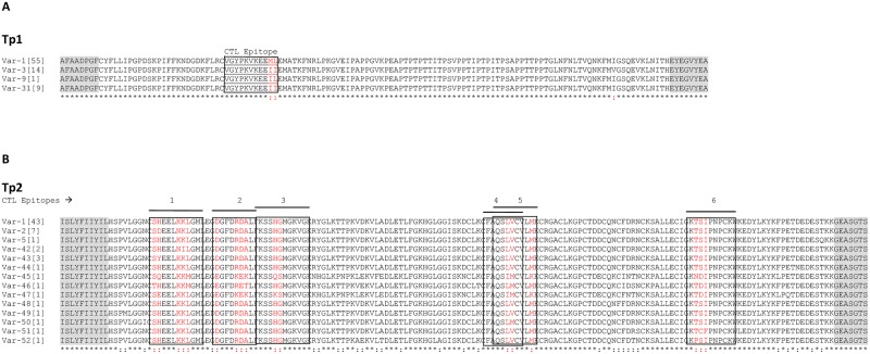
Fig 2. Multiple amino acid sequence alignment of Tp1 and Tp2 antigen variants present in cattle from South Sudan.
(A) Multiple sequence alignment of the four Tp1 antigen variants. Antigen variants Var-1, -3 and -9 were first described by Pelle et al. (2011) [17]. (B) Multiple sequence alignment of 14 Tp2 antigen variants. The naming of the antigen variants follows the nomenclature by Pelle et al. (2011) [17]. Antigen variants Var-1, -2 and -5 were first described by Pelle et al. (2011) [17]. The CD8+ T-cell target epitopes are boxed and the polymorphic residues in the epitopes are shown in red. The frequency of each variant amongst the samples tested is indicated in square brackets. Residues conserved in all sequences are identified below the alignment (*). The shaded flanking regions are equivalent to the positions of the secondary (nested) PCR primers, and are not included in estimations of the percentage of the residues that are conserved.

