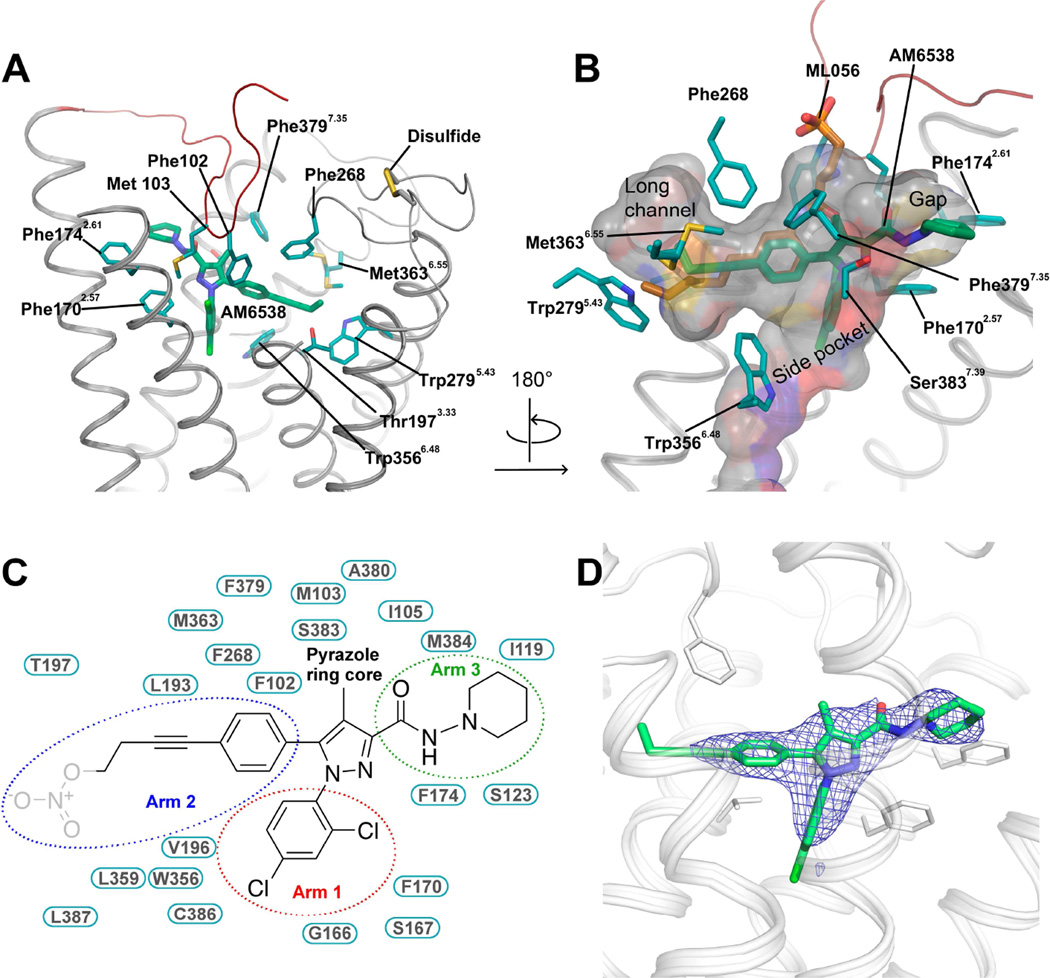
Figure 3. Analysis of the Ligand Binding Pocket of CB1.
(A) Key residues in CB1 for AM6538 binding. AM6538 (green carbons) and CB1 residues (teal carbons) involved in ligand binding are shown in stick representation. The receptor is shown in gray cartoon representation. (B) The shape of the ligand binding pocket. AM6538 (green carbons) and ML056 (brown carbons) are shown in stick representation. (C) Schematic representation of interactions between CB1 and AM6538. The 2,4-dichlorophenyl ring in the red circle is termed as arm 1; The 4-aliphatic chain substituted phenyl ring in the blue circle is termed as arm 2; The piperidin-1-ylcarbamoyl in the green circle is termed as arm 3. The nitrate group, which was not observed in the electron density, is shown in gray. (D) Electron density maps calculated from the refined structure of the CB1-AM6538 complex. |Fo|-|Fc| omit map (blue mesh) of the ligand AM6538 is shown (contoured at 3 σ). See also Figure S3.

