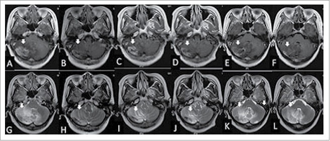
Figure 3.

The woman was diagnosed with radiation brain necrosis for 1 year. Gadolinium-enhanced T1-weighted MRI in February 2015 showed widespread scattered irregular enhancement (A) and T2-weighted FLAIR MRI showed a large edema in the surrounding tissue (G). After bevacizumab treatment (3.27 mg/kg) in February 2015, the volume of necrosis (B) and edema (H) was reduced. Fifteen weeks after bevacizumab administration, gadolinium-enhanced T1-weighted MRI showed that the volume of necrosis (C) was enlarged and T2-weighted FLAIR MRI showed the edema (I) in the surrounding tissue was enlarged in June 2015, hence bevacizumab treatment (3.27 mg/kg) was given for the second time. At the July 2015 follow-up, the volume of necrosis in gadolinium-enhanced T1-weighted MRI (B) and edema in T2-weighted MRI (J) was reduced significantly again. In the October 2015 follow-up, the volume of necrosis in gadolinium-enhanced T1-weighted MRI (E) and edema in T2-weighted FLAIR MRI (K) was enlarged and the neurological symptoms were aggravated again; thus the patient was treated with bevacizumab (3.27 mg/kg) for the third time. Eight weeks after the third treatment of bevacizumab, the volume of necrosis in gadolinium-enhanced T1-weighted MRI (F) and edema in T2-weighted MRI (L) was reduced again.
