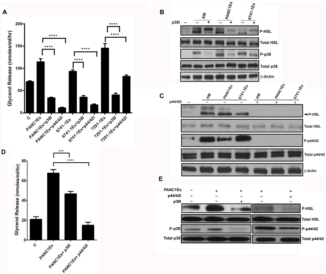
Figure 5. Lipolytic effect of AM is mediated through MAP kinases (p38 and ERK1/2) in subcutaneous adipocytes.
(A) Glycerol release in 3T3-L1 cells treated with PC-exosomes (PANC1Ex, 6741-1Ex & 7291-1Ex., 50 µg) and inhibitors of the MAPK pathway: p38 inhibitor (p38I; 10 µM), p44/42 inhibitor (p44/42I; 10 µM) and compared to exosomes only treatment group, C refers to untreated cells. ****, P<0.0001. B) Western blot showing phosphorylation of p38 and HSL in cells treated with p38I and AM (10 nM) or p38I and PC-exosomes (PANC1Ex & 6741-1Ex) for 24 hours. (C) Western blot showing phosphorylation of p44/42 and HSL in cells treated with p44/42I (10 µM) with AM peptide (10 nM), p44/42I with PC-exosomes (PANC1Ex & 6741-1Ex) and compared to AM, PANC1Ex and 6741-1Ex treated groups respectively. + and − refers to treatment with and without inhibitor. (D) Glycerol release in human adipocytes treated with p38 inhibitor (p38I; 20 µM) and p44/42 inhibitor (p44/42I; 10 µM) for 6 hours followed by incubation with PANC1Ex (50 µg) or treatment with PANC1Ex (50 µg) alone for 24 hours. (B) Western blot depicting phosphorylated levels of p38, p44/42, and HSL in human adipocytes treated with inhibitor (+) and without inhibitor in the presence (+) and absence (−) of PANC1 exosomes.

