
Figure 5. The crosstalk of p53 and NF-κB pathways regulated by Drp1-mediated mitochondrial fission was essential for cell cycle progression.
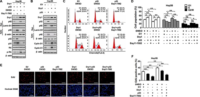
(A) Western blot analyses for protein levels of Drp1, p53, p21, Rb, phosphorylated-RB (p-Rb) in whole cells or p65 in cytoplasm and nucleus of Hep3B cells with treatment as indicated. (B) Western blot analyses for protein levels of Drp1, p53, cyclin D1 and cyclin E1 in whole cells or p65 in cytoplasm and nucleus of Hep3B cells transiently transfected with Drp1 and/or p53 expression vector as indicated. (C and D) Cell cycle analysis by flow cytometry in Hep3B cells 48 h after transfection with expression vector of Drp1 and/or p53. In the group with NF-κB inhibitor, cells were also treated with Bay11-7082 or DMSO (used as a control therapy) 12 h before analysis. (E and F) Cell proliferation was evaluated by EdU incorporation assay in Hep3B cells as indicated in Panel (C and D). Scale bar, 50 μm. The results shown are the mean ± SEM from three separate experiments.