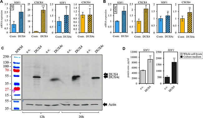
Figure 4. DUX4-transfected cells overexpress SDF1 and CXCR4 genes.

qRT-PCR analysis of the expression of CXCR4 and SDF1 mRNA in human immortalized myoblasts (MB) (A) and bone marrow mesenchymal stem cells (BSMC) (B) transiently transfected with DUX4- or, DUX4c-expressing plasmids or an empty vector (pCI-Neo). Average of three independent experiments is shown, error bar represent standard deviation (SD), t-test p-value < 0.05 (*). Gene expression level of the control sample normalized to GAPDH was set to 1. (C) Western blot analysis of protein lysates of human immortalized myoblasts (MB) transfected with DUX4 and DUX4c plasmids for 12 and 20 h. DUX4 (52-kDa) and DUX4c (47-kDa) proteins were stained with 9A12 antibody. (D) SDF1 protein concentration was measured using ELISA in the whole cell lysate (diluted 10-fold) or cell culture medium (concentrated 20 times) of human immortalized myoblasts (MB) 24 h after transfection with pCI-Neo-DUX4, or pCI-Neo (e.v.) plasmids. Average and standard deviation of 3 independent experiments are shown, t-test p-value < 0.05 (*).
