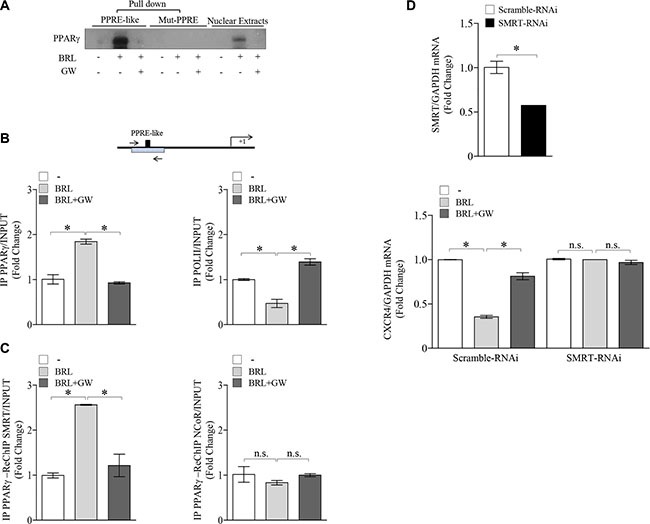
Figure 3. Ligand-activated PPARγ binds to a PPRE-like site within CXCR4 promoter.

(A) DAPA on nuclear extracts from MCF-7 cells treated with vehicle (−), BRL 10 μM with or without GW 10 μM for 3 h. PPRE-like or mutated (Mut-PPRE) biotinylated oligonucleotides were used. Nuclear Extracts, positive control. (B) Schematic representation (upper panel) of PPRE-like site in CXCR4 promoter region. Chromatin Immunoprecipitation (ChIP) assay (lower panel) with anti-PPARγ and anti-POL II antibodies in MCF-7 cells treated with vehicle (−), BRL 10 μM with or without GW 10 μM for 1 h. (C) ChIP with the anti-PPARγ antibody was re-immunoprecipitated (Re-ChIP) with the anti-SMRT or anti-NCOR antibodies. The CXCR4 promoter sequence including the putative PPRE site was detected by Real-time-PCR with specific primers (see Material and Method section). (D) mRNA levels of SMRT (upper panel) and CXCR4 (lower panel) evaluated by Real-time RT-PCR in MCF-7 cells transfected with control RNAi (Scramble RNAi) or SMRT RNAi for 24 h and then treated with vehicle (−), BRL 10 μM with or without GW 10 μM for 24 h as indicated. Each sample was normalized on its GAPDH mRNA content. The results are expressed as fold change respect to the vehicle-treated cells. The values represent the mean ± SD of three different experiments, each performed with triplicate samples. *P < 0.05. n.s. = not significant.
