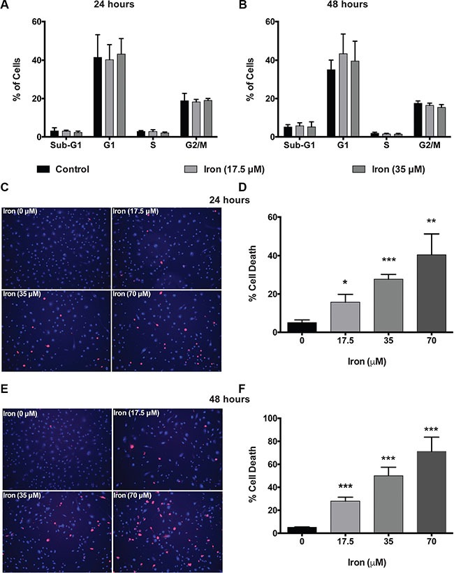
Figure 2. Cell-permeable iron induces cell death in VEGF-A stimulated endothelial cells.

Cell-cycle analyses of HUVEC-I treated with cell-permeable iron (17.5 μM and 35 μM) were carried out by flow cytometry. (A) Cells treated for 24 hours. (B) Cells treated for 48 hours. Data represent mean ± SD of four independent experiments. (C) Representative images of PI stained HUVEC-I stimulated with VEGF-A (100 ng/ml) in the presence of cell-permeable iron for 24 hours. Hoescht-33342 (blue, total cells) and PI (red)-stained (dead cells) are shown (10× magnification). (D) The histogram represents cell death analyses from three independent experiments. Cell death was calculated as a percentage of PI positive nuclei from the total number of Hoescht-33342 positive nuclei (blue) per field. Data represent mean ± SD. *P < 0.05, **P < 0.01, ***P < 0.001. (E) Representative images of HUVEC-I stimulated with VEGF-A (100 ng/ml) in the presence of cell-permeable iron for 48 hours. Dead cells were identified by PI staining. (F) The histogram represents cumulative data of cell death analyses from three independent experiments. Data represent mean ± SD. ***P < 0.001.
