
Figure 7. Cell-permeable iron inhibits VEGFR-2 signaling through FAK and p38 MAPK affecting migration.
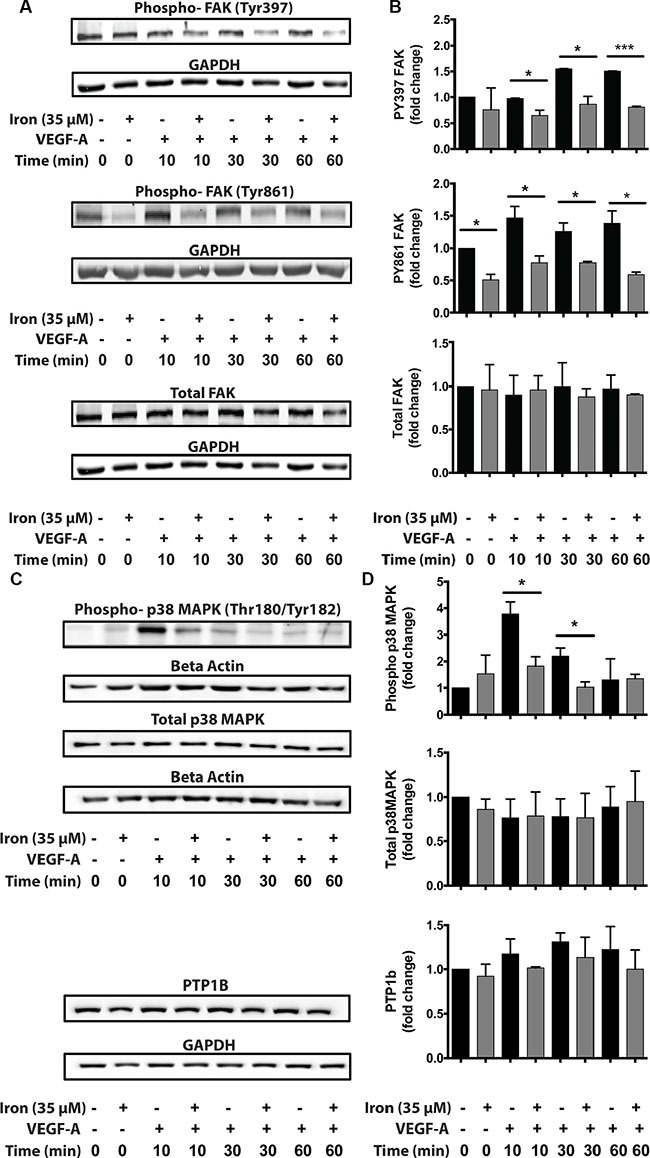
HUVEC-I cells were stimulated with VEGF-A (100 ng/ml) for 10 min to 60 min in the presence of 35 μM iron. (A) Cell lysates were then analyzed by Western blots to determine relative levels of phosphorylated FAK (pTyr-397 and pTyr-861). (B) Cumulative data showing relative levels of FAK phosphorylation and total FAK levels from two-four independent experiments. (C) Western blots showing phosphorylated p38-MAPK and total levels of PTP1B following VEGF-A stimulation of HUVEC-I cells in the presence of cell-permeable iron. (D) Densitometric analyses of Western blots from two independent experiments. Beta-Actin levels were used for normalization of phospho and total p38 MAPK levels. GAPDH levels were used to normalize PTP1B levels. *P < 0.05, ****P < 0.0001.