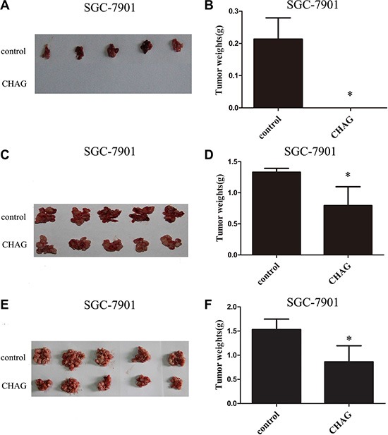
Figure 2. CHAG inhibits colonization and growth of gastric cancer cells in peritoneal cavity.

(A and B) The inhibition of CHAG on the colonization of gastric cancer cells in peritoneal cavity. Ten million SGC-7901 cells (suspended in 400 μl PBS) with or without CHAG (500 μg/ml) were injected into peritoneal cavity of nude mouse. Twenty five days later, the mice were executed, the tumors were excised, and the weights of the tumors of the different groups were calculated. (C and D) The inhibition of CHAG on the early growth of gastric cancer cells. Ten million SGC-7901 cells suspended in 400 μl PBS were injected into peritoneal cavity of nude mice. Two hours late, 400 μl CHAG solution (at concentration of 500 μg/ml) was injected into the cavity once only. The mice were fed normally for 8 weeks and then were executed and the tumors were collected and weighed. (E and F) The inhibition of CHAG on mid-term growth of gastric cancer cells. The cells were given to the mouse same as described in (C and D) Seven days later, 400 μl CHAG solution (at concentration of 500 μg/ml) was injected into peritoneal cavity of the mouse and the injection was repeated once a week. After 7 weeks, the mice were executed, the tumors were collected and weighed. A, C, and E were images of tumors from the mice in control and CHAG groups. B, D, and F were results of weight analysis of the tumors in corresponding group. The data shown were means ± SD. (*P < 0.01, compared with the control group).
